
Specimen Preparation - Electron Microscopy
On this page:
- Resin Embedding
- Critical Point Drying (CPD)
- Plunge-Freezing and Freeze-Drying
- Ultramicrotomy, Array Tomography, and Cryo-sectioning
- Sputter and Carbon Coating
- Glow Discharging
- Plasma Cleaning
Resin Embedding
The Electron Microscopy facility within the Microscopy Bioscience Platform provides a full TEM specimen preparation service for thin sectioning. This includes supply of fixatives (for immersion or perfusion fixation), staining, dehydration, and resin embedding. Commonly used resins include Quetol, LR White, and Araldite.
Processing protocols are tailored to the sample type and intended imaging or analysis. While we offer standard workflows, we are also able to accommodate specialised protocols, please consult with us early in your project to ensure the most suitable preparation approach.

Main specimen preparation lab
Critical Point Drying (CPD)
The Quorum critical point dryer enables safe drying of both small and relatively large biological or material samples for SEM. Biological specimens are typically fixed with chemical agents (e.g. formaldehyde, glutaraldehyde, osmium tetroxide), then washed in deionised water and dehydrated, commonly using ethanol. Some materials can be processed unfixed.
Large samples (e.g. whole insects, tissue sections, organoids, or coverslip-grown cells) can be placed directly in the specimen boat. Smaller specimens (e.g. tiny organisms, hydrogel particles) are enclosed in microporous capsules to prevent loss during processing. Inside the CPD chamber, samples are flushed with liquid CO₂, then dried by carefully raising the temperature and pressure past the critical point. Once dried, they are ready for mounting and sputter-coating.

Quorum E3100 critical point drier with large specimen chamber (left). Metal specimen boats (dimensions: length ~ 75 mm, diameter ~ 12 mm) are available for drying larger samples and microporous specimen capsules are used as boat inserts for enclosing small samples (right).
Plunge-Freezing and Freeze-Drying
Plunge-freezing followed by freeze-drying is a rapid method for preparing fixed or unfixed SEM samples. Specimens are first rinsed in cold deionised water to remove buffer or media salts, then plunged into liquid ethane cooled by liquid nitrogen (LN₂). Once vitrified, samples can be fractured under LN₂ to expose internal structures.
Frozen specimens are mounted in LN₂-cooled brass inserts and transferred to a turbo freeze dryer, typically left overnight. After drying, they are ready for mounting and sputter-coating. This method is best suited to thin samples, as freezing rates decline with depth, increasing ice crystal formation and structural damage. Slightly larger samples can be used if the area of interest is near the surface. Drying capacity depends on insert type: up to 6 larger or 8 flat (e.g. coverslip) samples per run.
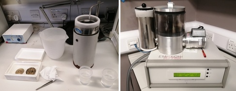
Manual setup for plunge-freezing in liquid ethane (left) and sample transfer via liquid nitrogen-cooled brass inserts to a LN2-cooled freeze-drier (right; Quorum/Emitech K775X).
Ultramicrotomy, Array Tomography, and Cryo-sectioning
Ultramicrotomy
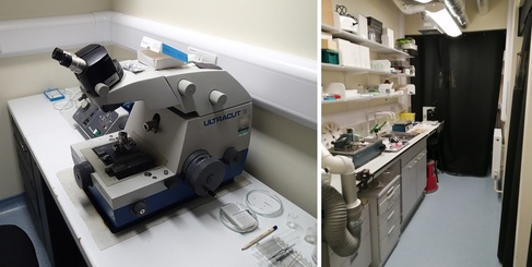
Our EM facility houses three Leica Ultracut ultramicrotomes for preparing ultrathin sections for electron microscopy. Two are reserved for staff use; one is available to trained users during office hours. We provide glass knives, hotplates, methylene blue, and mounting medium for thick section preparation. Users must supply their own diamond knives for ultrathin sectioning.
Array Tomography – ATUMtome
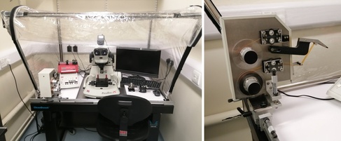
We also host an RMC Boeckeler ATUMtome, on loan from Dr Cahir O’Kane (Department of Genetics, University of Cambridge). This system combines an ultramicrotome with an automated tape-collecting reel that captures serial thin sections onto a film ribbon. These are mounted on 4-inch silicon wafers and imaged on the Verios SEM in backscatter mode. Image stacks can be computationally reconstructed into 3D volumes. Access is available by consultation only.
Cryo-sectioning – Leica CM3050 S Cryostat
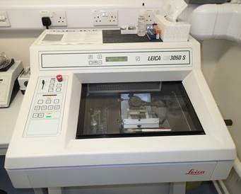
Our cryostat enables sectioning of frozen tissue samples for histology or fluorescence imaging. Section thickness ranges from 0.5 to 300 µm, with typical use between 5–15 µm. Samples are embedded using non-reactive compounds such as OCT (Scigen) or Leica Tissue Freezing Medium. Users must bring their own disposable blades and slides. Booking is available to trained users during office hours.
Sputter and Carbon Coating
We use a Quorum Emitech metal sputter coater with gold or iridium targets to coat SEM specimens prior to imaging, improving conductivity and image quality. For EDX analysis or array tomography, a Quorum carbon coater, using braided carbon cord, is used to deposit conductive carbon films. This system is suitable for SEM samples, TEM grids, and large silicon wafers (up to 4 inches). Both coaters operate with an argon gas supply.
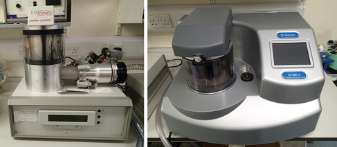
Quorum Emitech K575X metal sputter coater (left) and Quorum Q150T E carbon coater (right).
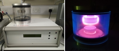
Glow Discharging
We use a Quorum Emitech K100X glow discharger with argon gas for treating TEM grids, glass coverslips, and other substrates. A standard protocol involves a negative glow discharge at 25 mA for up to 2 minutes. This process renders hydrophobic carbon films more hydrophilic, promoting even spreading of biological macromolecules—particularly ahead of negative staining.
Plasma Cleaning
The Fischione 1020 single-port plasma cleaner uses a 27% oxygen/argon gas mix and can be used to clean TEM specimen rods and TEM or SEM samples prior to imaging. It is also suitable for cleaning glass substrates for use in high-resolution fluorescence microscopy techniques.
