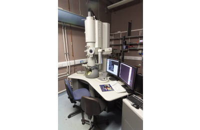
The Electron Microscopy Team offer a wide range of specimen preparation techniques for both scanning electron microscopy (SEM) and transmission electron microscopy (TEM) samples as a full service, as well as facilitating access for experienced users to our Tecnai G2 TEM and Verios 460 SEM. We are also happy to discuss any aspect of experimental design, sample preparation and to assist in interpretation of results where our expertise allows.
Research Technical Professionals in our Platforms and Facilities provide essential scientific support, and we follow the Royal Microscopy Society guidelines for fair attribution, ensuring their contributions are routinely acknowledged in publications, with authorship considered for substantial intellectual input. See our Fair Attribution Policy on the Platform overview page for more information.
Contact
For any electron microscopy enquiries, please contact us: electron-microscopy@bio.cam.ac.uk
Book a microscope and request training on our booking system PPMS >>>
Electron Microscopy Team
Dr Filomena Gallo
Electron Microscopy Technical Specialist
fg337 [at] cam [dot] ac [dot] uk
Training and Booking
We use Stratocore Pasteur Platform Management System (PPMS) to manage equipment bookings and training.
As a new electron microscopy user, we ask that you:
- create a PPMS account with your CRSid (if you have one).
- create a project. You are required to enter the grant code in the “account number” box. Account number format: XXXX/### e.g. ABCD/123.
- Email electron-microscopy@bio.cam.ac.uk to discuss your project and arrange the necessary training and services you require
The Electron Microscopy platform team will then discuss sample and imaging requirements with you. We are very happy to discuss and provide advice on experimental design.
After training, please book to use electron microscopes through our PPMS booking system.
Equipment
| PPMS System Name | Tecnai G2 TEM | Verios 460 SEM |
| Department (Room) | PDN (Anatomy Building, 20) | PDN (Anatomy Building, 21) |
| System Image | ||
| Voltages | 80, 120 and 200 keV |
Accelerating voltage range of 1-30 keV, plus low voltage range of 350 – 1000 eV |
| Emission source | LaB6 | Field-emission gun |
| Stage | compu-stage with ± 60 degree stage tilt for 3D tomography | Computer-driven, high precision stage with 360 degree continuous rotation and +60 degree to -10 degree stage tilt |
| Detectors/Cameras | bottom-mounted AMT CCD camera |
Everhart-Thornley (ETD) and Through-Lens detectors (TLD) for routine SE- and BSE-imaging in field-free mode (large field of view/depth of field) and immersion-mode (high resolution imaging) Concentric backscatter detector (CBS) for differential detection of BS electrons scattered at various angles from the beam axis; custom-mode allows selection of individual detector segments Segmented STEM detector (STEM III) for TEM grid samples with custom settings for bright-field (BF), dark-field (DF) and high-angle annular dark field (HAADF) imaging Energy-dispersive X-ray detector (Ametek window-less silicon drift detector) for elemental analysis running with EDAX Genesis or TEAM software Mirror (MD) and In-column (ICD) detectors allowing detection of BS electrons for increasingly high Z-contrast imaging |
| Capabilities |
High angle annular darkfield STEM (HAADF-STEM) for Z-contrast imaging. Elemental analysis by EDX (Peltier-cooled Ametek silicone drift detector; Genesis/ Team software) Two single tilt holders, one low background EDX holder, one tomography holder |
Large range of image resolution and scan speed settings; custom settings for interlace scanning, multiple image integration and drift correction to improve image acquisition even of charging samples; definition of scan presets for easy and quick sample navigation and image acquisition UniColor mode (UC mode, monochromator, selectable below 5 keV/25 pA) to improve image quality at low keV/probe current settings Energy-dispersive X-ray detector (Ametek window-less silicon drift detector) for elemental analysis running with EDAX Genesis or TEAM software. Cryo-SEM: Quorum cryo-transfer system PP3010T for loading, coating and imaging quick-slush frozen, hydrated specimens Beam-deceleration mode in room temperature and cryo-mode MAPS software for automated acquisition of high resolution images from large areas of SEM specimens or TEM blockfaces |
Specimen Preparation
The electron microscopy team supports a number of specimen preparation methods for both Scanning and Transmission Electron Microscopy and houses a diverse range of equipment.
Techniques supported include:
-
Resin Embedding
-
Critical Point Drying
-
Plunge-freezing/ and freeze-drying
-
Ultramicrotomy, Array Tomography, and Cryo-sectioning
-
Sputter and carbon coating
-
Glow Discharging
-
Plasma Cleaning
Please contact us to discuss the details of your requirements.
Other services available in the School of Biological Sciences
Electron microscopy | Cambridge Stem Cell Institute
The Cambridge Stem Cell Institute maintains both Transmission Electron Microscopy and Scanning Electron Microscopy on the Cambridge Biomedical Campus and is open to University of Cambridge and external users.
Microscopy | Sainsbury Laboratory
The Sainsbury Laboratory maintains a cryo-scanning electron microscopy capability in central Cambridge that supports a variety of plant science research and is open to University of Cambridge and external users.


