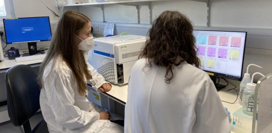
Flow cytometry is a powerful tool that provides rapid analysis of cells in solution through optical signalling and light detection. Flow cytometry is most widely used in mammalian biology, however the technique can be applied to almost any particle with useful optical properties including bacteria, viruses, subcellular organelles, and artificial particles, for example, polystyrene beads bearing ligands or antibodies.
Flow cytometry has applications in immunology, molecular biology, bacteriology, virology, cancer biology and infectious disease monitoring.
School-based Facilities open to all include:
-
Flow Cytometry at the Department of Pathology
-
Flow Cytometry at the Department of Veterinary Medicine
Flow Cytometry at the Department of Pathology
Instruments:
Cell Sorters
- BD FACS Discover S8 with CellView technology
- BD Aria III
- BD Aria IIu
Analysers
- Two Aurora Spectral Analysers (Aurora A and Aurora B)
- CytoFLEX LX
- CytoFLEX S
- Attune NxT
- LSR II
Location
The facility is based in the Department of Pathology (see map)
Contact
Flow Cytometry Facility Manager: Joana Cerveira
Email: flow.cytometry@path.cam.ac.uk
Access and Training
The Flow Cytometry Facility is open to all members of the University and also to commercial users from biotech companies. In house training provided.
More information and booking
Flow Cytometry at the Department of Veterinary Medicine
Equipment
- BD LSR Fortessa 18 colour flow cytometry analyzer
- Imagestream Mark I Imaging Flow Cytometer
- Exoview - Exosome Imager
- Zeiss LSM780 Confocal Microscope
- Leica CM1950 Cryostat for the cutting of frozen sections
Location
The facility is based in the Department of Veterinary Medicine (see map)
Contact
Imaging Facility Manager: Rachel Hewitt
Email: microscopy@vet.cam.ac.uk
Access and Training
The flow cytometry facility is open to all members of the University, external academic institutions and industry. In house training is provided.
More information and booking

