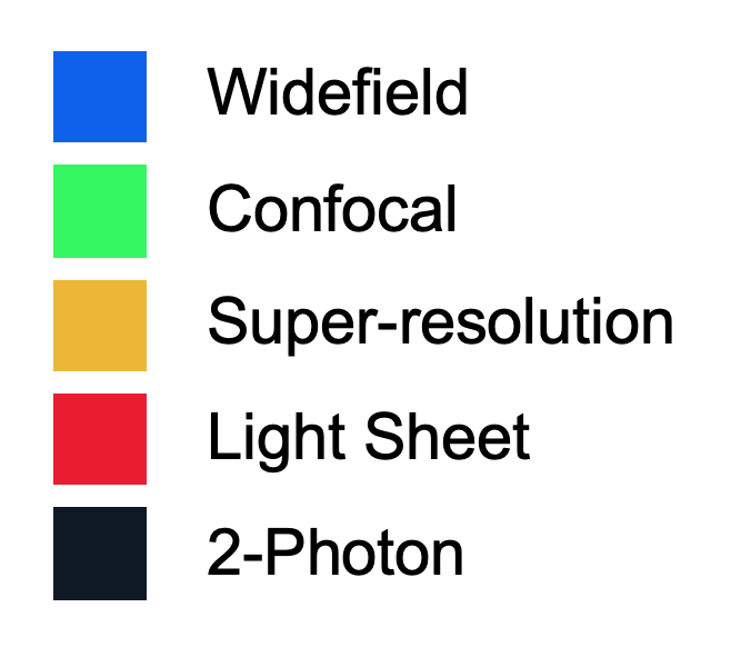
Light Microscopy technologies available in the School of Biological Sciences include widefield and confocal light microscopes as well as more complex light sheet and super resolution imaging modalities for which we provide dedicated support to users.
The Light Microscopy team also provide a wide range of other advanced applications and modules, including Fluorescence Recovery After Photobleaching (FRAP), Fluorescence Resonance Energy Transfer (FRET), TauSense Technology Imaging Tools (analogous to Fluorescence Lifetime Imaging Microscopy (FLIM)) and AiryScan and offers live imaging under biological containment level 2 (CL 2) conditions.
We also provide training and support for the diverse range of microscopes available across Downing, New Museums and Old Addenbrooke's sites in the centre of Cambridge.
If you are unsure which technique or microscope best fits your needs, please contact us on light-microscopy@bio.cam.ac.uk and we can provide guidance and examples from previous similar projects.
Research Technical Professionals in our Platforms and Facilities provide essential scientific support, and we follow the Royal Microscopy Society guidelines for fair attribution, ensuring their contributions are routinely acknowledged in publications, with authorship considered for substantial intellectual input. See our Fair Attribution Policy on the Platform overview page for more information.
Contact
For any light microscopy enquiries, please contact us: light-microscopy@bio.cam.ac.uk
Book a microscope and request training on our booking system PPMS >
Light Microscopy Team
Dr Antonina J. Kruppa
Biological Microscopy Coordinator
ajk62 [at] cam [dot] ac [dot] uk
Dr Martin Lenz
Advanced Light Microscopy
Senior Technology Developer
mol21 [at] cam [dot] ac [dot] uk
Light Microscopy Drop-in Clinics in the School of the Biological Sciences
We hold regular clinics during office hours in Departments. Members of the University are welcome to drop by and chat to us about any bioimaging questions that you may have. If you are an external user, please contact us: light-microscopy@bio.cam.ac.uk
Current sessions:
Mondays, 13:30 - 15:00
- Department of Physiology, Development and Neuroscience, Anatomy Building, The Wolfson Light Microscopy Laboratory
- Department of Zoology, Main Building, Room T13B
Tuesdays
- 13:30 - 15:00 - Department of Pathology, Tennis Court Road Building, Room 318
- 15:00 - 16:30 - Department of Psychology, Downing site, Psych Sanctuary
Wednesdays
- 9:30 - 11:00 - Department of Biochemistry, Sanger Building, Tennis Court Road, table in atrium
- 11:00 - 12:30 - Department of Pharmacology, Tennis Court Road, breakout space Level 1
Thursdays
- 13: 30 - 15:00 - Department of Plant Sciences, Botany Building, Tea Room
- 15:00 - 16:30 - Department of Genetics, Downing site, Tea Room
Training and Booking
Please only request training if you have a sample ready that you have prepared, or contact us for sample preparation advice and suggestions.
We use Stratocore Pasteur Platform Management System (PPMS) to manage equipment bookings and training.
As a new microscopy user, we ask that you:
- create a PPMS account with your CRSid (if you have one).
- create a project. You are required to enter the grant code in the “account number” box. Account number format: XXXX/### e.g. ABCD/123.
- select “request a training” on the PPMS homepage and complete the Light Microscopy Training Request form.
The Light Microscopy platform team will then discuss sample and imaging requirements with you and schedule a training session on the most suitable microscope for your needs. We are very happy to discuss and provide advice on experimental design.
After training, please book to use light microscopes through our PPMS booking system.
Requesting an Assisted Session
Please email us first (light-microscopy@bio.cam.ac.uk) to arrange a suitable time when we can assist you.
Booking Guidelines
-
5 hour limit per day (during normal working hours).
-
5 hour limit per calendar week (during normal working hours).
-
A 'last minute' booking made within 5 hours of present time are excluded from the above limits.
-
After 5 hours of continuous imaging the price drops by 50% per hour.
-
After 10 hours of continuous imaging the price drops by a further 50% per hour.
-
If the needs of your project require special consideration please contact us.
Microscope Specifications and Locations
-
Widefield microscopes
-
Confocal microscopes
-
Light Sheet microscopes
-
Super-resolution microscopes
-
Two-photon microscopes
Our microscopes are located on the Downing and New Museum sites
Other services available in the School of Biological Sciences
Imaging | Cambridge Stem Cell Institute
The Cambridge Stem Cell Institute maintains a suite of light microscopes at the Cambridge Biomedical Campus that are open to all members of Cambridge University and in house training is provided.
Cellular Imaging and Analysis Facility | Department of Veterinary Medicine Research Facilities
The Department of Veterinary Medicine maintains a cellular imaging and analysis facility in West Cambridge that includes light microscopy and is open to both University of Cambridge and external users.
Microscopy | Sainsbury Laboratory
The Sainsbury Laboratory maintains a large core facility with a suite of 23 individual microscopes and a wealth of experience and expertise in imaging plant tissues. This facility is open to both University of Cambridge and external users.
Gurdon Institute Imaging Facility Website
The Gurdon Institute maintains a wide variety of advanced light microscope systems and provides support with experimental design, sample preparation, image acquisition, computational image analysis and data presentation as well as providing comprehensive training on all their microscopes.

