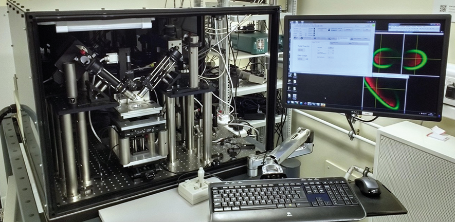
Our custom-built light sheet microscope, funded by the Wellcome Trust, was developed from ground up towards the requirements of our users. The upright geometry allows for easy sample mounting inside a standard petri dish as often required for long time-lapse whole organism imaging. Since different organisms need different imaging conditions the sample mount can be temperature controlled.
The operating software is based on Labview. A home build software package allows flexible adaption to experimental requirements, while a clean and intuitive user interface has been developed for the rather complex instrument.
Located in the Department of Physiology, Development and Neuroscience, Anatomy building, room 22A
Technical Specifications and Capabilities
- Standard excitation wavelengths (445nm, 488nm, 515nm, 561nm, 638nm) in combination with a number of emission filters are available to cover a broad range of fluorophores.
- Hamamatsu Orca Flash 4 camera with up to 50 planes/second
- Adaption of excitation volume (light sheet length) to different samples with tunable lenses
- Heated sample holder
Example Applications
Single-colour time-lapse imaging of zebrafish retina over 48 hours
The movie shows the temporal activation of Notch signalling across the retina from about 24hpf until 72hpf, using the TP1:VenusPEST transgenic zebrafish line. This zebrafish line contains a Notch responsive element (TP1) coupled to a Venus fluorophore which has been destabilized with a PEST sequence for fast degradation. During the movie one can see how Notch is first activated throughout the progenitor population and eventually becomes restricted within the Mueller glial cells.
The imaging was performed on our custom built upright light sheet microscope, using 515 nm excitation and acquiring volumes of ~300 μm x 300 μm x 200 μm every 2.5 minutes. Shown are maximum intensity projections in three directions.
These images were taken in collaboration with Xana Almeida and Julia Oswald, Harris group.
Comparison between 2-Photon Excitation and 1- Photon excitation light-sheet imaging of zebrafish eye
The movie/ images show a 72hpf Ath5:gapRFP transgenic zebrafish retina. In this transgenic, the Ath5 promotor drives the expression of membrane tagged RFP in retinal ganglion cells as well as in photoreceptors, horizontal and amacrine cells. We can observe the axons of retinal ganglion cells exiting the eye and projecting towards the brain. These images were taken in collaboration with Xana Almeida, Harris group.


