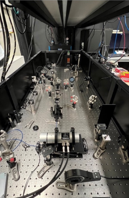
This Wellcome Trust-funded project is developing a versatile photo-manipulation system that combines holographic optical tweezers, patterned illumination, and photoablation with real-time confocal microscopy. The system is designed to ablate biospecimens, control their behaviour, and quantitatively characterise their properties while simultaneously imaging the sample with a high-end confocal microscope.
Located in the Department of Physiology, Development and Neuroscience, Anatomy building, Optical Development Lab
This project aims to develop a versatile photo-manipulation system to ablate biospecimens, control their behaviours and characterise their properties quantitatively whilst imaging the sample with a high-end confocal microscope.
Capabilities
- The complete system is composed of:
- holographic optical tweezers
- a bespoke Digital Micromirror Device (DMD) based patterned illumination unit
- a Ti:Sapphire laser based ablation unit integrated on a commercial Nikon TiE2 frame.
- The holographic optical tweezers use a NIR laser to trap microspheres for measuring cell’s properties.
- The patterned illumination unit offers high resolution (1920 x 1200) and high speed for photo-activation or photo-conversion of biospecimens.
- The ablation unit emits femtosecond laser with the maximum peak power of 300kW for cutting cell membranes or ablating large-scale tissues.
- The microscope adapts 5 objectives including:
- 10x, 20x, 40x (Silicon oil), 60x (Oil) and 100x (Oil), for imaging various biospecimens from single cell level to the entire embryo level.
- The system can acquire two-colour live confocal imaging of biospecimens excited by 405/405/445/470/520/528/555/640 nm lasers.
Technical Specifications
Nikon Ti2 microscope frame
- Objective-10X; 20X; 40X (Silicon oil); 60X (Oil); 100X (Oil)
- Temperature control
CrestOptics X-light spinning disk confocal
- Spinning disk - 50 μm pinhole / 250 μm spacing or 50 μm pinhole / 500 μm spacing
- Excitation lasers - 405; 445; 470; 520; 528; 555; 640 nm
- Camera – 2x sCMOS Prime95B
- Simultaneous dual colour imaging
Optical Tweezers
- Trapping laser – 1064 nm CW laser
- Calibrated stiffness range - 5 pN/μm to 200 pN/μm
- Μulti-spot trapping capability
Patterned Illumination
- DMD based pattered illumination, (1920 x1200 pixel)
- Activation light source - 405; 445; 470; 520; 528; 555; 640 nm
- Simultaneous activation/deactivation of different areas
Applications
Various studies have been undertaken based on this system with the collaboration with biologists, including:
- membrane tension measurements on HeLa cells,
- photo-activation and photo-deactivation of zebrafish embryos,
- photo-conversion of fly embryos,
- large scale ablation on zebrafish tissues,
- membrane cutting on fly embryos
Furthermore, our system has the capability to undertake complex bio-experiments without transferring specimens between different microscopes. To illustrate, imaging of live HeLa cells, quantifying the cell membrane tension, studying cell shape governed by actin-mCherry, locally photoactivating a single cell, photo-ablating the membrane of the cell and tracing the variation of membrane tension.


