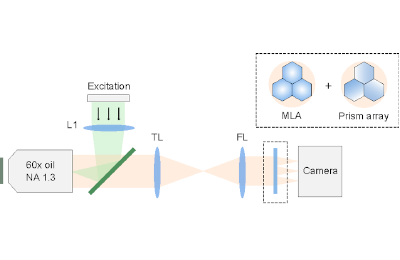
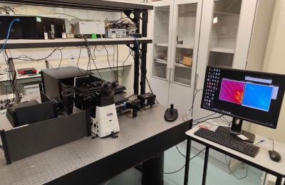
Vortex Light Field Microscopy (VLFM) is a novel technique for 3D spectral single-molecule imaging that allows the nanoscale structures in cells to be imaged with high spatial and spectral precision. By adding an azimuthally oriented prism array to our previous single molecule light field microscopy, VLFM achieves simultaneous spatial and spectral localization on a single detector.

Located in the Department of Physiology, Development and Neuroscience, Anatomy building, Optical Development Lab
Technical Specifications
- Excitation lasers available at 405, 488, 561, 638 nm. The setup works with a broadband emission between 400-800 nm.
- Dichroic: Chroma, ZT405/488/561/640rpcv2
- Filters: Semrock, NF03-405/488/561/635E; Semrock, BLP01-647R-25
- Hamamatsu Flash 4.0 V2 (pixel size 6.5 μm) as well as a Photometrics Prime95B (pixel size 11 μm)
- HCImageLive (included with the purchase of a Hamamatsu camera)
- Pixel size in sample space = 184 nm
Capabilities
- Simultaneous 3D and spectral localisation, enabling multicolour 3D single particle tracking.
- 25 nm spatial and 3 nm spectral precision over 4 μm depth-of-field.
- Small PSF footprint, allowing for higher imaging density than PSF engineering methods (such as double-helix and tetrapods).
- No registration between channels, as we only use a single detector.
Applications
- With DNA-PAINT for highly multiplexed super-resolution imaging.
- With smFISH for high-throughput RNA imaging
- With nano-barcoding for spatial proteomics study in neurons.
- Colocalisation analysis with or without FRET
- Monitoring the spectral fluctuations or environmental sensitivity of fluorescent probes.


